Figures 56-64

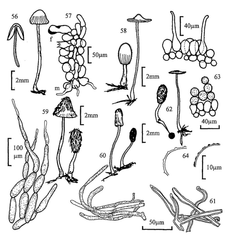
Fig. 56. Habit sketch of a stipitate agaric, Psathyrella stercoraria, with section.
Fig. 57. Sketch of gill section of Psathyrella sp., showing position of marginal (m) and facial (f) cystidia.
Fig. 58. Coprinus pellucidus, habit and vertical section of cap cuticle.
Fig. 59. C. pseudoradiatus, habit and veil constituents.
Fig. 60. C. vermiculifer, habit and veil constituents.
Fig. 61. C. filamentifer, veil constituents.
Fig. 62. C. stercoreus, habit.
Fig. 63. C. cordisporus, vertical section of cap showing nature of veil cells on the cap cuticle.
Fig. 64. Veil cells with structural (l) and superficial crystalline (r) ornamentation.

22(21) Cap without setules. 23
- Cap with setules. 24
23(22) Cap minute, 1-5mm high before expanding, reddish orange at first, soon fading. Basidiospores almost globose to triangular in one view, elliptic in another, 7-10 × 7-9 × 5.5-6.5µm. (2- and 4-spored forms have been found).
Coprinus miser
- Cap larger, up to 15mm when expanded. Basidiospores pip-shaped, 7.5-8.5 × 9.5-11 × 9.5-11.5µm. (4-spored).
Coprinus nudiceps
24(22) Spores hexagonal, 10-13 × 6.5-7.5µm. Cap purplish.
Coprinus hexagonosporus
- Spores ellipsoid. Cap brown or reddish, without purplish tints. 25
25(24) Basidia 4-spored. 26
- Basidia 2-spored. Spores 11-13 × 5.5-7µm. Facial cystidia absent.
Coprinus bisporus
(Coprinus sassii, not yet recorded in British Isles, has 2-spored basidia with very large ellipsoid spores up to 20µm long).
26(25) Cap with a mixture of hyaline and brown thick-walled setules. Spores 9-10 × 5.5-6µm, with eccentric germ pore. Facial cystidia absent.
Coprinus heterosetulosus
- Cap with only one type of setule. Facial cystidia present or absent. 27
27(26) Facial cystidia present. Spores 7.9-13.3 × 4.4-6.4µm, with apical germ pore.
Coprinus stellatus
- Facial cystidia absent. Spores elongate and narrow, rarely greater than 5µm wide, with apical germ pore. Fruit body usually quite small, up to 6mm before expanding.
Coprinus pellucidus (fig. 58)
(Several species in the group, e.g. C. congregatus and C. ephemerus have been found on straw/dung mixtures).
28(21) Veil strongly adhering to cap. Spores elliptic ovate, 15-20 × 8-12µm. Stem with distinct ring. Usually on buried dung.
Coprinus sterquilinus
- Veil more floccose or powdery. Stem lacking ring or, if present (C. ephemeroides), fruit body small with 5-angled spores less than 10µm long. 29
29(28) Veil composed of filamentous units. 30
- Filamentous units, if present, masked by a preponderance of rounded cells. 35
30(29) Veil composed of strings of sausage-shaped, thin-walled, hyaline cells. 31
- Veil composed of rather narrow, slightly thickened hyphae. 32
31(30) Spores large, 11-14 × 6-7µm. Cap up to 1cm before expanding. Fruit body with or without a rooting base.
Coprinus radiatus
- Spores smaller, up to 9µm long. Cap up to 6mm before expanding. Fruit body without a rooting base.
Coprinus pseudoradiatus (fig. 59)
(C. cinereus is found on straw/dung mixture and C. macrocephalus, with large spores, has been recorded on raw dung).
32(30) Veil citrus- or lime-yellow, or a mixture of hyaline and brown strongly coloured hyphae. 33
- Veil grey or whitish. 34
33(32) Veil of yellow hyphae. Spores 10.5-12.5 × 6-7.5µm.
Coprinus luteocephalus
- Veil with brown hyphae. Spores 7-9 × 3.5-5µm.
Coprinus poliomallus
34(32) Veil hyphae thin-walled. Spores 6.5-7.5 × 5µm, 'shouldered' about the apiculus.
Coprinus filamentifer (fig. 61)
- Veil hyphae thin- and thick-walled, often with clamps. Spores elliptic-oblong, 9-10 × 5-6µm.
Coprinus vermiculifer (fig. 60)
(Coprinus flocculosus, with spores 11.5-16.5 × 6-9.5µm, can be found on straw/dung mixtures).
35(29) Stem with small, distinct ring. Spores subglobose to lentiform and 5-angled, 6-9 × 6.5-8 × 5-6µm.
Coprinus ephemeroides
- Stem at most with fibrils, even then rarely forming a faint ring zone. 36
36(35) with setules in addition to veil. 37
- Cap without setules. 38
37(36) Cap cystidia tapered. Spores 11-14 × 5-6.5µm.
Coprinus heptemerus
-
Cap cystidia capitate. Spores 10-11 × 6-7µm. Coprinus curtus
38(36) Veil of inflated bladder-like cells attached to filamentous units. Spores 7.5-8 × 4.5-5.5µm.
Coprinus utrifer
- Veil of globose and subglobose cells and filamentous units often encrusted or with minute projections found sometimes at cap margin. 39
39(38) Globose cells, if ornamented then possessing crystalline or amorphous material (dissolved by 1N HCl, fig. 64.) 40
- Globose cells covered in small fine blunt projections on the walls (not removed by 1N HCl, fig. 64). 45
40(39) Basidia 2-spored. 41
- Basidia 4-spored. 42
41(40)
Spores 14-17 × 8.5-10 × 12.5-14µm. Coprinus pachyspermus
- Spores smaller, 9-11 × 6-6.5 × 8-9µm.
Coprinus cordisporus (2-spored form)
42(40) Spores less than 10µm long.
Coprinus cordisporus (fig. 63)
(C. patouillardii is known on garden refuse, and an undescribed species with lemon-shaped spores has recently been found).
- Spores 10µm or more long. 43
43(42) Veil soon discolouring greyish, drab or buff, Spores 11.5-14.5 × 6-8 × 7.5-9µm.
Coprinus cothurnatus
- Veil remaining snowy white, only slowly discolouring greyish. 44
44(43) Fruit bodies several cm tall. Spores 15-19 × 8.5-11.5 × 11-13µm.
Coprinus niveus
- Cap small, 5-6mm at first. Spores 14-16 x 8-9 × 10-12.5µm.
Coprinus latisporus
45(39) Basidia 3-spored. 46
- Basidia 4-spored. 47
46(45)
Spores narrow, 8.5-11 × 5-6.2µm. Coprinus triplex
- Spores broad, 9-10 x 6-6.5 × 6-7µm, slightly flattened in face view.
Coprinus trisporus
(These are possibly a single taxon).
47(45) Spores 7-8 × 4-4.5µm, perispore not visible in water or alkali mounts.
Coprinus stercoreus (fig. 62)
- Spores 9µm or more long. 48
48(47) Spores 9-11 × 5.5-6µm. Perisporal sac none or incomplete or indistinct.
Coprinus foetidellus
- Spores longer, 10.8-13.5 × 5.5-7µm, with distinct perispore with dark lines and inclusions. Distinctive smell of gas.
Coprinus narcoticus
(C. sclerotiger is found on straw/dung mixtures, and the smaller C. tuberosus on garden refuse etc.).
49(20) Spores not discoloured in conc. H2SO4. 50
- Spores discolouring in conc. H2SO4. Gills not spotted at maturity. 66
50(49) Cap cuticle cellular. Gills spotted at maturity. (More often on rich, 'dungy', soils. P. subbalteatus, with copper coloured cap, drying paler but retaining a dark marginal zone, occurs in gardens on mulch etc.).
(Panaeolus) 51
- Cap cuticle filamentous. 56
51(50) Velar remnants very obvious, either as an appendiculate veil or as a distinct ring. 52
- Lacking all velar remnants. 54
52(51) Cap distinctly pigmented, with appendiculate veil. 53
- Cap pale coloured, smooth, semi-globate, soon cracking. Gills with marginal cystidia only.
Panaeolus papilionaceus
53(52) Cap brown, smooth, sometimes viscid, not exceedingly wrinkled.
Panaeolus campanulatus
- Cap grey, olivaceous, even black, with contrasting white appendiculate veil.
Panaeolus sphinctrinus
54(51) Cap with or without appendiculate veil, but always with distinct ring.
Panaeolus semiovatus
- Cap lacking veil. 55
55(54) Cap pinkish ochraceous to tawny-buff. Lacking facial cystidia.
Panaeolus speciosus
- Cap whitish or slightly yellowish. With facial cystidia.
Panaeolus antillarum
56(50) Gills with facial cystidia often containing yellow amorphous material when seen in ammonia solution or deep blue with cotton blue.
(Stropharia) 57
(Blue-green S. cyanea & S. aeruginosa often occur in rich garden soils).
- Gills lacking facial cystidia. Never with yellowing cystidia in ammonia.
(Psilocybe) 58
(Red-capped P. aurantia can be found on straw/mulch mixtures in gardens).
57(56) Cap sticky, semi-globate ± expanding at maturity. On raw dung.
Stropharia semiglobata
- Cap plano-convex, often broad with a central umbo, margin flaring with age. On dungy mixtures in gardens.
Stropharia stercoraria
58(56) Stipe bluing, with ring. Spores ellipsoid, 11-14 × 6.5-7.5µm. Fruit body with mealy smell and taste.
Psilocybe fimetaria
- Stipe lacking distinct ring, or if with ring or ring zone 2-spored and/or stem not bluing. Fruit body without mealy smell and taste. 59
59(58) Stem always with distinct ring. Basidia 2-spored. Spores 15-20µm long.
Psilocybe luteonitens
- Stem with or without ring. Basidia 4-spored. If with ring, spores smaller. 60
60(59) With ring zone. 61
- Lacking velar remnants on stem, or only appendiculate teeth at cap margin. 62
61(60) Spores slightly angular/limoniform, 11-13(14) × 7-8µm. Often on sewage sludge.
Psilocybe merdaria
-
Spores 13-14 × 7.5-8.5µm. Psilocybe moelleri
62(60)
Spores 14-20 × 8-10µm. Psilocybe subcoprophila
- Spores smaller. 63
63(62) Spores lentiform, angled, 6-8(8.5) × 4.5-5.5 × 3.75-4.5µm.
Psilocybe bullacea
(P. crobula, occasional on dung, differs in lacking purple colour in gills, and slightly smaller, ovoid, not angular, spores).
- Spores larger. 64
64(63)
Spores ellipsoid to slightly amygdaliform. Psilocybe merdicola
- Spores lentiform, angular. 65
65(64)
Spores 11-13(14) × 7-8(9)µm. see Psilocybe merdaria, 61
-
Spores 12-15 × 8-9.5µm. Psilocybe coprophila
66(49) Round cells on cap as a micaceous veil. (Re-examine gill face; if different sized basidia and facial cystidia separating the gills are present go to Coprinus at 21).
Psathyrella sphaerocystis
- Cap lacking veil, or if present then fibrillar. 67
67(66) White copious veil at margin or also covering cap centre. Spores 10-12 × 5.5-6µm.
Psathyrella coprobia
- Lacking copious veil. 68
68(67) With red edge to gill. Spores 12-13 × 6-6.5µm, with central germ pore.
Psathyrella stercoraria
- Lacking red gill edge. Spores with eccentric germ pore.
Psathyrella coprophila
(P. fimetaria differs in spore size; there are several members of the P. prona group which grow on soil/straw mixtures).
69(2) Fruit body club-shaped.
Typhula setipes (fig. 65)
(Clavaria acuta often grows on peaty soil in pots in greenhouses).
- Fruit bodies effuse, resupinate 70
70(69) Fruit-body cobweb-like and greyish white. Basal hyphae 3-4.5µm wide. Spores sub-globose, 4.5µm diam. (Generally on old dung or straw/soil mixtures).
Athelia coprophila
(If with spiny spores 5-6µm diam., see the recently recorded Tomentellopsis echinospora).
- Fruit-body with pores, white or flushed slightly ochraceous, brownish or greyish. (On clods of soil in dunged land).
Cristella candidissima
71(1) Fruit body either a cup containing several 'eggs' or a single orange or yellowish gelatinous sphere. 72
- Fruit-body effuse, without distinct shape. 73
72(71) Fruit-body whitish or pale yellow, up to 2.5mm diam., splitting at maturity to shoot away the orange/yellow spore mass.
Sphaerobolus stellatus (fig. 66)
- Fruit-body cup shaped, with silvery-grey 'eggs'. (Usually on dung and straw or attached to rabbit pellets).
Cyathus stercoreus
(Cyathus vernicosus often grows in plant pots on rich soil).
73(71) Basidia with transverse septa. Spores 11 × 7µm. Fruit body pinkish.
Platygloea fimicola
(Not British; included for completeness. Pilacrella solani, with a glistening stipitate head, has been isolated from dungy soil).
- Basidia with longitudinal septa. Spores 14-18 × 9-10µm. Fruit body cream-white or ivory.
Sebacina incrustans
Figures 65-67

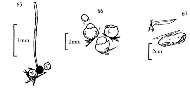
Fig. 65. Habit sketch of Typhula sp. Note attachment to sclerotium.
Fig. 66. Sphaerobolus stellatus, habit.
Fig. 67. Clitopilus passackerianus, a sessile agaric—habit sketch and section.

Key 4. Zygomycota

1 Spores formed in multispored sporangia (figs 68, 70, 72, 75, 76) or in few-spored sporangioles (figs 70, 73). 2
- Multispored sporangia and globose sporangioles absent. Spores formed singly on terminal, lateral or intermediate vesicles (figs 74, 79, 80, 82-86), or in short chains (figs 77, 78, 81). 11
2(1) Sporangiophore stout, simple, with a subsporangial swelling and a basal swelling buried in the substrate. Sporangia tough walled, black, projected some distance towards the light when mature, and sticking to whatever they hit.
Pilobolus (fig. 76)
e.g. spores pale yellow, 8-10 × 5-6µm - P. crystallinus
spores orange, 12-20 × 6-10µm. - P. kleinii
- Sporangiophores not stout; sporangia not violently discharged. 3
3(2) Sporangial wall black, tough, not readily broken when touched. Sporangia with a sticky base, becoming attached to whatever they contact after the marked elongation of the white sporangiophores at maturity.
Pilaira (fig. 75)
e.g. spores yellowish, 8-10 × 6µm - P. anomala
spores colourless, 11-13 × 6-8µm - P. moreaui
- Sporangial wall diffluent, spores readily removed in a droplet, or fragile and then spores easily dispersed by external violence. 4
4(3) Sporangiophores stiff and metallic in appearance, growing towards the light and often to great length (5-30cm).
Phycomyces
e.g. spores 10.5-30 × 6.5-17µm; columella pyriform; sporangiophores up to 30cm - P. nitens
spores 8-13 × 5-7.5µm; columella spherical or ovoid; sporangiophores up to 30cm - P. blakesleeanus
- Sporangiophores white, not reaching extreme lengths. 5
5(4) Small lateral sporangia (sporangioles) present. 10
- Sporangioles absent. 6
6(5) Sporangiophores usually grouped, less often single, connected by stolon-like hyphae. 7
- Sporangiophores arising singly, or if grouped then lacking stolon-like hyphae. 9
7(6) Stolons joining groups of sporangiophores often with rhizoids at the base of the group. 8
- Sporangiophores arising singly or in groups from stolons, which may be 'rooted' at intervals along their length, but rarely beneath the groups of sporangiophores.
Absidia (fig. 71)
e.g. sporangiophores grouped, rhizoids poorly developed; spores 2.5-4.5µm diam. - A. corymbifera
sporangiophores grouped, rhizoids strongly developed; spores 2.5-3.5µm diam. - A. orchidis
8(7) Sporangiophores mostly unbranched.
Rhizopus (fig. 69)
e.g. spores irregularly angular-ovoid, 8-14 × 11µm - R. nigricans
- Sporangiophores with a whorl of branches beneath the main sporangium, each with a small columellate sporangium. Spores 6-8.5µm.
Actinomucor elegans