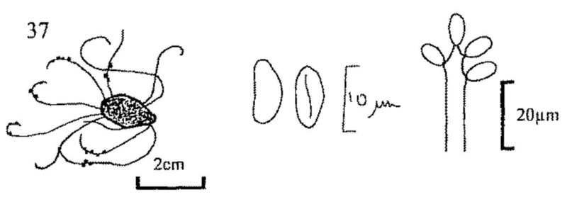
Fig. 37. Wawelia sp., stromatic filaments with perithecia growing from a rabbit pellet, ascospores, and conidiophore and conidia.

Fig. 37. Wawelia sp., stromatic filaments with perithecia growing from a rabbit pellet, ascospores, and conidiophore and conidia.
| 146(141) | Asci 8-spored. Spores 20-31 × 9.5-15µm. | |||
| Bombardioidea bombardioides | ||||
| - | Asci 4-spored. | 147 | ||
| 147(146) | Spores 24-34 × 15-19(20)µm. Basal germ pore less distinct than the apical one. | |||
| Bombardioidea serignanensis | ||||
| - | Spores 34-43 × 16-22µm. Distinct germ pore at each end of spore. | |||
| Bombardioidea stercoris | ||||
|
148 (key 1,1) |
Fruit bodies solitary or in small groups, each a subglobose, fertile, light brown head on a slender sterile stalk. Head soon bursting to expose the yellow ochraceous spore mass. On mixtures of bird droppings, cast pellets and decaying animal material. | 149 | ||
| - | Fruit bodies superficial, lacking a distinct stalk. | 150 | ||
| 149(148) | Spores 5-8 × 2-3µm. Head 1-2mm diam. | |||
| Onygena corvina (fig. 38) | ||||
| - |
|
|||
| 150(148) | Fruit bodies with an external wall of loosely anastomosing and interwoven hyphae, and with ± specialised terminal cells (gymnothecia, fig. 39). | 151 | ||
| - | Fruit bodies with a well defined parenchymatic wall (cleistothecia, fig. 46). | 161 | ||
| 151(150) | Gymnothecia with simple thin-walled, ± uniform and poorly developed hyphae constituting the outer hyphal sheath. | 152 | ||
| - | Gymnothecia with thick-walled hyphae modified at their ends into appendages, or if thin-walled then always accompanied by appendages (i.e. curled, toothed or pointed hyphae). | 155 | ||
| 152(151) | Gymnothecia red-orange to brick-red. Ascospores orange, subglobose to ellipsoid, with an equatorial furrow, smooth, 4.5-5.5 × 3.5-4.5µm. | |
| Arachniotus ruber (fig. 40) | ||
| - | Gymnothecia white or yellow, never orange or brick-red. Ascospores without an equatorial furrow. | 153 |
| 153(152) | Gymnothecia white. Ascospores hyaline, ellipsoid, smooth, 3-4 × 2-2.5µm. | |
| Arachniotus candidus | ||
| - | Gymnothecia distinctly pigmented, yellow or brown. Ascospores larger than 4µm. | 154 |
| 154(153) | Gymnothecia yellow brown. Ascospores orange to brownish, slightly lenticular, smooth or slightly roughened, 5-6.5 × 3.3-4.6µm. | |
| Arachniotus confluens | ||
| - | Gymnothecia lemon yellow. Ascospores lemon yellow, lenticular, smooth, 5-6 × 3-4.5µm. | |
| Arachniotus citrinus | ||

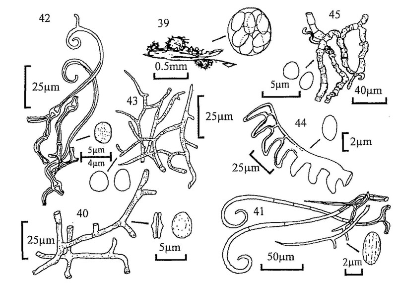
Fig. 39. Habit sketch of a gymnothecium and ascus.
Figs 40-45. Spores and peridial hyphae.
Fig. 40. Arachniotus ruber. Fig. 41. Myxotrichum chartarum.
Fig. 42. Gymnoascus californiensis. Fig. 43. Gymnoascus reesii.
Fig. 44. Ctenomyces serratus. Fig. 45. Arthroderma curreyi.
| 155(151) | Gymnothecia possessing only thick pigmented hyphae. | 156 |
| - | Gymnothecia possessing ± thin, hyaline hyphae with only a few, although often distinctive, appendages (i.e. comb-shaped end cells or dumb-bell shaped asperulate cells accompanying twisted and bent hyphae). | 160 |
| 156(155) | Gymnothecia brown-black or dark greenish-grey, with external hyphae with spine-like branches and septate, hooked appendages. Ascospores orange brownish, ovate, delicately striate, 4-5.2 × 2.4-3.3µm. | |
| Myxotrichum chartarum (fig. 41) | ||
| - | Gymnothecia never black, and, if possessing thick-walled hyphae, then appendages never septate. Ascospores smooth, or if ornamented then asperulate or echinulate. | 157 |
| 157(156) | Gymnothecia rose to orange-brown or yellowish. Appendages curved or irregularly branched and pointed, never verticillately branched. Ascospores smooth, or at most asperulate. | 158 |
| - | Gymnothecia red-brown with appendages verticillately branched. Ascospores 3-4.5 × 2-2.8µm, yellowish brown, lenticular. | |
| Actinodendron verticillatum | ||
| 158(157) | Gymnothecia rosy pink when young, becoming browner, with spines and curved, non-septate hairs. Ascospores hyaline, globose to subglobose, asperulate, 3-5 × 2.5-4µm. | |
| Gymnoascus californiensis (fig. 42) | ||
| - | Gymnothecia yellow. Ascospores smooth. | 159 |
| 159(158) | Gymnothecia yellow to yellow-brown, without elongated appendages but with thick-walled branches, few of which are pointed. Ascospores globose-ellipsoid, yellow to brownish, 3-4.5 × 3.5µm. | |
| Gymnoascus reesii (fig. 43) | ||
| - | Gymnothecia golden yellow to reddish-brown, with acute-ended appendages. Ascospores lenticular, smooth, hyaline, 2.5-3.5 × 2-2.5µm. | |
| Pseudogymnoascus roseus | ||
| 160(155) | Gymnothecia orange brown, with comb-like appendages. Ascospores slightly lenticular, pale orange, 3.3-3.6 × 2-2.6µm. | |
| Ctenomyces serratus (fig. 44) | ||
| - | Gymnothecia whitish to pale ochraceous, particularly when dry, with few appendages but those present twisted and bent, and their branches constricted with regular or irregular dumb-bell shaped cells. Hyphal walls asperulate or with protuberances. Ascospores smooth, lenticular, hyaline, 2.4-3.3 × 2µm. | |
| Arthroderma curreyi (fig. 45) | ||
| 161(150) | Asci relatively large, 100-200-spored, 1-3/fruit body. 'Cleistothecia' minute, <100 (rarely <250)µm diam., immersed. | |
| see Thelebolus etc. (Key 1, 86) | ||
| - | Asci with 8 or fewer spores. | 162 |
| 162(161) | Ascospores purple at maturity, large, 50-70 × 25-35µm, epispore with a few longitudinal cracks. | |
| see Ascobolus immersus (Key 1, 48) | ||
| - | Ascospores smaller, hyaline, yellow, olivaceous, brown or black. | 163 |
| 163(162) | Ascospores olivaceous, brown or black, at least in part. | 164 |
| - | Ascospores aseptate, hyaline, yellow or other pale colours. | 174 |
| 164(163) | Ascospores 4-celled (cf. Sporormiella), with germ slits, readily fragmenting. Asci clavate, bitunicate. Cleistothecia black, shiny, up to 500µm diam. | 165 |
| - | Ascospores 1- or 2-celled. | 166 |
| 165(164) | Ascus stalk up to 20µm long. Ascospores 25-32 × 5µm. | |
| Preussia vulgaris | ||
| - | Ascus stalk 30-60µm long. Ascospores 26-38 × 5-7µm. | |
| Preussia funiculata (fig. 47) | ||
| 166(164) | Ascospores 2-celled. | 167 |
| - | Ascospores 1-celled | 170 |
| 167(166) | Spores unequally 2-celled, one brown ellipsoid, with an apical germ pore, 10-12 × 6.5-7.5µm, the other a basal hyaline, cylindrical pedicel, 6-8 × 3µm. Cleistothecia black, globose, up to 250µm diam., covered with flexuous brown hairs up to 1mm long. Asci evanescent. | |
| Zopfiella erostrata | ||
| - | Spores equally 2-celled. | 168 |
| 168(167) | Spores not constricted at the septum, ellipsoid, golden-brown, 25-30 × 10-15µm with 1-3 guttules in each cell. Cleistothecia gregarious on a mycelial mat, whitish to pale orange, up to 500µm diam. | |
| Heleococcum aurantiacum (fig. 48) | ||
| - | Spores hyaline, divided into two almost globose cells by the constricting septum. Ascomata superficial, globose, dark coloured. | |
| (Mycoarachis) 169 | ||
| 169(168) | Asci 8-spored, 5.5-11µm diam. Spores 5-5.5 × 3-3.5µm. | |
| Mycoarachis inversa | ||
| - | Asci 4-spored, 6-6.5µm diam. Spores 4.5-5 × 2-2.5µm. | |
| Mycoarachis tetraspora | ||
| 170(166) | Asci broad-clavate, (1)-2-(3)-spored, 30-50 × 13-18µm. Spores brown-black with short ridges and warts, subglobose, 12-15.5 × 11-12.5µm, with a single germ pore. | |
| Copromyces bisporus (fig. 49) | ||
| - | Asci 8-spored. | 171 |
| 171(170) | Spores globose, sooty brown, 3µm diam. Cleistothecia gregarious, with basal spirally coiled appendages, black, 100-200µm diam., partially immersed in a white to red felty hyphal mat. | |
| Pleuroascus nicholsonii | ||
| - | Spores larger, ellipsoid or limoniform. | 172 |

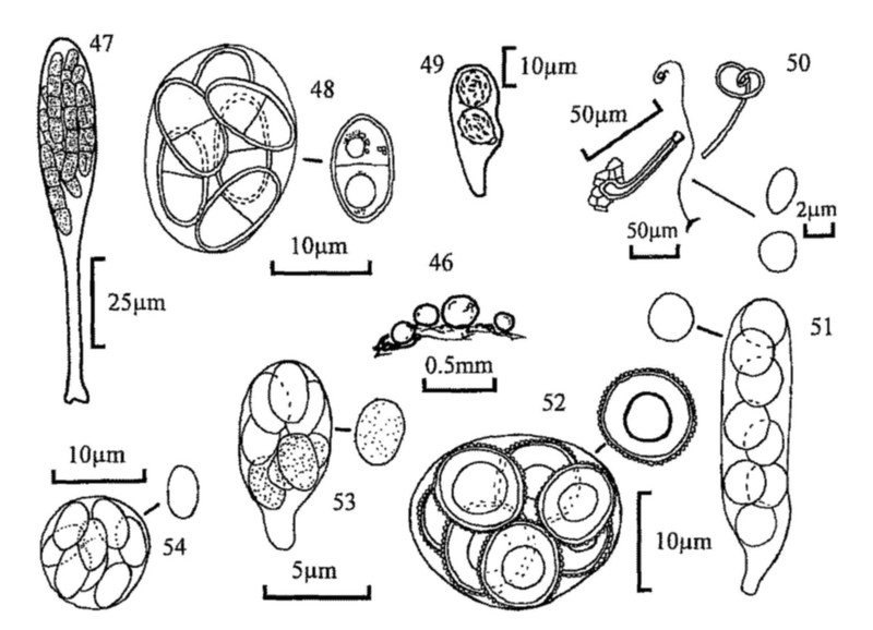
Fig. 46. Habit sketch of cleistothecia.
Figs 47-54. Asci and spores.
Fig. 47. Preussia funiculata. Fig. 48. Heleococcum aurantiacum.
Fig. 49. Copromyces bisporus. Fig. 50. Arachnomyces nitidus.
Fig. 51. Orbicula parietina. Fig. 52. Roumegueriella rufula.
Fig. 53. Aphanoascus stercoraria. Fig. 54. Pseudeurotium ovale.
| 172(171) | Spores olivaceous, limoniform, usually with an apical germ pore. Perithecia greyish or greenish, abundantly hairy, branched or simple, straight or curly. Asci pedicellate, soon disappearing. | |
| see Chaetomium at 5 | ||
| - | Spores darker, with 1 or more minute germ pores. Cleistothecia distinctly but not abundantly hairy. | 173 |
| 173(172) | Spores smoky brown, broadly ovoid, 9-14 × 6-9µm. Cleistothecial hairs short, up to 30µm. | |
| Thielavia wareingii | ||
| - | Spores dark brown, flattened limoniform, 13-16 × 10-13 × 8-9µm. Cleistothecial hairs of two types, some smooth, dark brown, arising from the base up to 3mm long, others greyish green, rough, up to ca 120µm. | |
| Thielavia fimeti | ||
| 174(163) | Cleistothecia produced within a common arachnoid mycelial mass. Spores smooth or minutely asperulate, yellow to yellow-brown, broadly ellipsoid, 4-5 × 3-5µm. | |
| Aphanoascus fulvescens | ||
| - | Cleistothecia single or gregarious, but not on or in a mycelial mass. | 175 |
| 175(174) | Cleistothecia 170-750µm diam., covered with long (several mm when extended), thick-walled, aseptate, helical appendages. Asci clavate cylindrical, evanescent, 35-62 × 12-21µm. Spores ellipsoid, hyaline, 12-17 × 9-12µm. | |
| Lasiobolidium spirale | ||
| - | Cleistothecia without coiled appendages. | 176 |
| 176(175) | Cleistothecia with hairs or appendages. | 177 |
| - | Cleistothecia smooth. | 178 |
| 177(176) | Cleistothecia black, shining, 100-200µm diam., with dark brown-black thick-walled hairs with hooked tips. Asci 8-15µm diam. Spores straw or copper coloured, ellipsoid, 4-7 × 3.5-4.5µm with de Bary bubble and a germ pore at each end. | |
| Kernia nitida | ||
| - | Cleistothecia reddish brown, less than 1mm diam., with long simple appendages curled at the tips. Spores hyaline, oblate, 3.55 × 2-3µm. | |
| Arachnomyces nitidus (fig. 50) | ||
| 178(176) | Ascospores globose, larger than 9µm. | 179 |
| - | Ascospores ellipsoid, up to 9µm. Asci always subglobose. | 180 |
| 179(178) | Ascospores, smooth, 9-13µm. | |
| Orbicula parietina (fig. 51) | ||
| - | Ascospores ornamented, 13-24µm Asci subglobose. Cleistothecia ochraceous, becoming yellowish brown or flushed cinnamon. | |
| Roumegueriella rufula (fig. 52) | ||
| 180(178) | Ascospores hyaline, then faintly yellowish, minutely spiny, 2.5-3 × 2-2.5µm. Cleistothecia pale, then dark brown. | |
| Aphanoascus stercoraria (fig. 53) | ||
| - | Ascospores hyaline, then brown, smooth, 5.5-6 × 3.5-4µm. Cleistothecia dark brown from the beginning. | |
| Pseudeurotium ovale (fig. 54) | ||
| 1 | Basidia single-celled (fig. 55). | 2 |
| - | Basidia transversely or longitudinally septate (fig. 55), or difficult to observe. | 71 |
| 2(1) | Fruit body agaricoid, i.e. mushroom-shaped with gills underneath cap (figs 56, 67). | 3 |
| - | Fruit body not agaricoid, without gills (figs 65, 66). | 69 |
| 3(2) | Spore print white or pale coloured, hyaline s.m. (Usually on straw/dung mixtures, never on raw dung except when very old). | 5 |
| - | Spore print coloured. | 4 |
| 4(3) | Spore print pinkish or pale cinnamon, honey-coloured s.m. (Usually on straw/dung mixtures, never on raw dung). | 6 |
| - | Spore print darker, in shades of brown or black. | 8 |
| 5(3) | Stem eccentric. Fruit body pure white. Spores ellipsoid, smooth. | |
| Pleurotellus s. lato | ||
| (If gills pink and spores longitudinally ridged see Clitopilus passackerianus, fig. 67) | ||
| - | Stem central. | 7 |

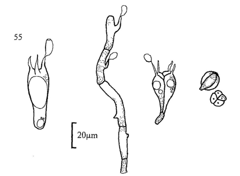
Fig. 55. From left, sketches of holobasidium, with mature basidiospore showing germ pore; auriculariaceous basidium; tremellaceous basidium, lateral view and as often seen in sections.
| 6(4) | Fruit body white, ivory or very pale tan, with a smell of cucumber. Gills decurrent. | |||
| Clitocybe augeana | ||||
| - | Fruit body yellow, with scaly cap. Gills free or just adnate. Fruit body with distinct ring and granular veil. (Commonly in plant pots. Probably associated with peaty material more than dung). | |||
| Leucocoprinus birnbaumii | ||||
| (L. cepaestipes and L. lilacinogranulosus occur in similar situations). | ||||
| 7(5) | Fruit body with amethyst/purple shades, with eccentric stem. Spores subglobose, slightly ornamented to nearly smooth. (On compost heaps in gardens). | |||
| Lepista nuda | ||||
| - | Fruit body with pink gills and distinct volva at stem base. Cap white to pale hazel. Stem white. Spores broadly ellipsoid, smooth. | |||
| Volvariella speciosa | ||||
| 8(4) | Spore print distinctly brown (fulvous, tawny, rust coloured etc.). | 9 | ||
| - | Spore print some darker shade, fuscous, fuliginous or violaceous black. | 20 | ||
| 9(8) |
|
|||
| (Has been found on straw/dung mixtures, never on raw dung). | ||||
| - | Stem lacking a veil. | 10 | ||
| 10(9) | Cap rich chrome yellow, viscid, soon reduced to a sticky mass, easily collapsing. | |||
| Bolbitius vitellinus | ||||
| - | Cap in shades of brown, never brightly coloured and if collapsing then cap elongate-cylindric and white to pale cream. | 11 | ||
| 11(10) | Spore print dull, sepia or snuff-brown. On rabbit pellets in sand dunes. | |||
| Agrocybe subpediades | ||||
| - | Spore print, brighter coloured, orange/rust brown. | |||
| (Conocybe) 12 | ||||
| 12(11) | Gill edge with irregularly fusoid cystidia with obtuse apices (lageniform). Cap viscid. | |||
| Conocybe coprophila | ||||
| - | Gill edge with distinctly capitate cells resembling a glass stoppered bottle (lecythiform). Cap never viscid, often pubescent under a lens. | 13 | ||
| 13(12) | Stem covered in long hairs. | 14 | ||
| - | Stem covered in lecythiform cells similar to those on gill edge, giving a farinaceous appearance under a lens. NEVER with long hairs. (Dung/straw mixtures). Large as in a Cortinarius. Spores smooth. | |||
| Conocybe intrusa | ||||
| (C. leucopus has been found on manured soil in gardens; C. antipus has hexagonal spores and grows on dung piles). | ||||
| 14(13) |
Stem with both long hairs and lecythiform cystidia. 
|
15 | ||
| - |
Stem with hairs and lageniform cystidia. 
|
16 | ||
| 15(14) | Spores 11-14 × 7-9µm. Taste and smell strong, of fresh meal. | |||
| Conocybe farinacea | ||||
| - | Spores large, over 15 × up to 10µm. Taste and smell none or slightly acidic. | |||
| Conocybe pubescens | ||||
| (C. subpubescens might be found on straw/dung mixtures, and differs in spores 11-13 × 6-8µm). | ||||
| 16(14) |
|
|||
| - | Basidia 4-spored. | 17 | ||
| 17(16) | Spores ellipsoid. | 18 | ||
| - |
|
|||
| 18(17) | Cap grey, contrasting with yellowish cream gills and pale stem. Spores 10.5-12.5 × 6-7µm. | |||
| Conocybe murinacea | ||||
| - | Cap pinkish brown or tawny. | 19 | ||
| 19(18) | Spores 11-12 × 7.2-7.8µm. Cap sienna. On raw dung. | |||
| Conocybe fimetaria | ||||
| - | Spores 10-12 × 6-7µm. Cap pinkish to cinnamon brown. On manured soil or sewage sludge. | |||
| Conocybe fuscomarginata | ||||
| (Conocybe siennophylla might be found on straw/dung mixtures or in soil in greenhouses. It differs in having smaller spores). | ||||
| 20(8) | Cap deliquescing to some degree at maturity. Basidia of 2 or 3 different sizes. | |||
| (Coprinus) 21 | ||||
| - | Cap not deliquescing. Basidia of one size only. | 49 | ||
| 21(20) | Veil on cap absent, cap either covered with small hairs (setules) or naked. | 22 | ||
| - | Cap covered with a granular, micaceous, powdery or fibrillar veil. | 28 | ||